IDIOPATIC GRANULOMATOUS MASTITIS: RADIOLOGICAL AND PATHOLOGICAL FINDINGS
GANİME DİLEK EMLİK, FAHRİYE KILINÇ, NECDET POYRAZ, Fatma Nur Çokbakar, Şeyma Ünüvar
- Year : 2025
- Vol : 5
- Issue : 3
- Page :
124-130
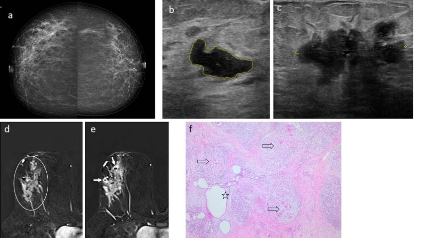
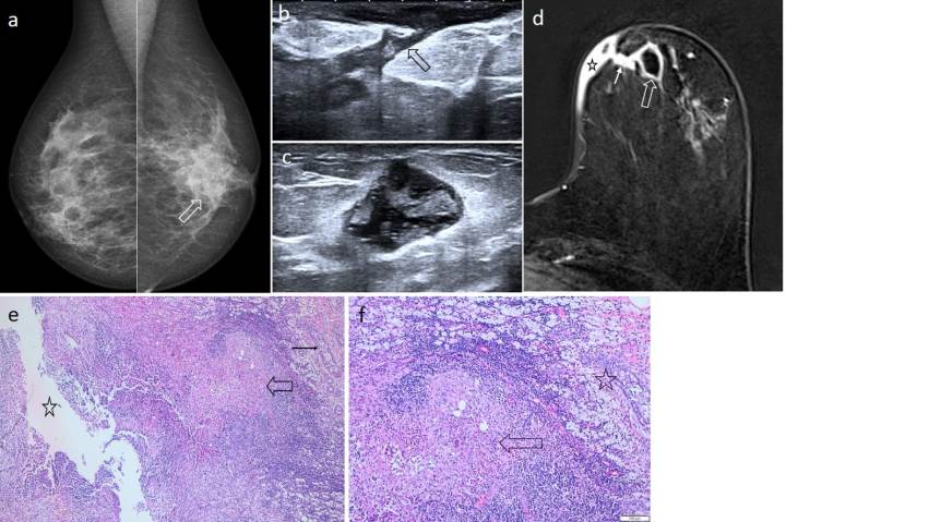
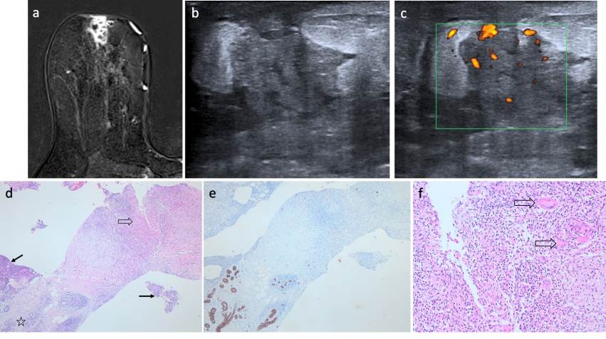
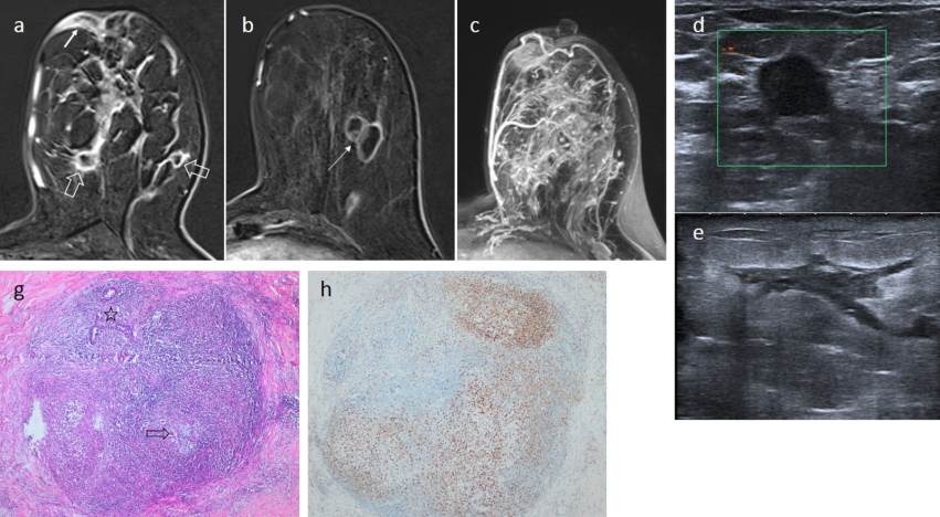
Idiopathic granulomatous mastitis is a rare, chronic, benign inflammatory disease of the breast that tends to be unilateral. It differs from other granulomatous breast diseases in that it lacks a definitive underlying etiology. However, lactation, hormonal changes, trauma, infections, and autoimmunity have been implicated in the etiology. Idiopathic granulomatous mastitis can mimic a wide spectrum of benign and malignant conditions, including bacterial mastitis, sarcoidosis, tuberculosis, vasculitic diseases, diabetic mastopathy, and inflammatory breast cancer; therefore, accurate diagnosis requires clinical, radiological, and pathological correlation. The diagnosis of idiopathic granulomatous mastitis is more often a diagnosis of exclusion. A woman of reproductive age, who has undergone lactation, and presents with a palpable breast mass and concomitant inflammatory findings (such as pain, fever, erythema) is the most common presentation. Radiological findings can be evaluated together with histopathological findings to arrive at a correct diagnosis. The radiological findings of idiopathic granulomatous mastitis can be nonspecific. However, radiological findings are quite helpful in establishing a preliminary diagnosis, determining the extent of the disease, and distinguishing it from malignant lesions. On mammography, the most common findings are an irregularly bordered mass or an asymmetrically increased opacity; on ultrasonography, hypoechoic tubular structures and an accompanying irregularly bordered hypoechoic mass; and on magnetic resonance imaging, the most common findings are segmentally distributed non-mass-like enhancing areas, a heterogeneous enhancing mass, and lesions representing a well-circumscribed, rim-like enhancing abscess. If idiopathic granulomatous mastitis is suspected clinically and radiologically, histopathological confirmation can be used to support the preliminary diagnosis. Currently, core needle biopsy is often preferred due to its less invasive nature and high diagnostic yield compared to excision. Histopathologically, it is seen as an inflammation involving the breast lobules and the fatty tissue surrounding the lobules, with sterile granulomas accompanied by Langerhans-type giant cells and epithelioid histiocytes.
Cite this Article As :
Description :
Yazarların hiçbiri, bu makalede bahsedilen herhangi bir ürün,
aygıt veya ilaç ile ilgili maddi çıkar ilişkisine sahip değildir. Araştırma,
herhangi bir dış organizasyon tarafından desteklenmedi.Yazarlar çalışmanın
birincil verilerine tam erişim izni vermek ve derginin talep ettiği takdirde
verileri incelemesine izin vermeyi kabul etmektedirler.
None of the authors, any product mentioned in this article,
does not have a material interest in the device or drug. Research,
not supported by any external organization.
grant full access to the primary data and, if requested by the magazine
they agree to allow the examination of data.
IDIOPATIC GRANULOMATOUS MASTITIS: RADIOLOGICAL AND PATHOLOGICAL FINDINGS, Review Article,
2025,
Vol.
5
(3)
Received : 13.08.2025,
Accepted : 18.11.2025
,
Published Online : 12.12.2025
Mevlana Tıp Bilimleri
ISSN: ;
E-ISSN: 2757-976X ;